Post Concussion Syndrome 
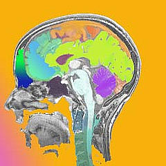
On November 21, 2012, brain imaging shows changes in the brains of people with post-concussion syndrome (PCS), according to a study published online in the journal Radiology.
PCS affects approximately 20 percent to 30 percent of people who suffer mild traumatic brain injury (MTBI) otherwise known as a concussion — defined by the World Health Organization as a traumatic event causing brief loss of consciousness and/or transient memory dysfunction or disorientation.
Symptoms of PCS include headache, poor concentration and memory difficulty. Conventional neuroimaging cannot distinguish which MTBI patients will develop PCS. “Conventional imaging with CT or MRI is pretty much normal in MTBI patients, even though some go on to develop symptoms, including severe cognitive problems,” said Yulin Ge, M.D., associate professor, Department of Radiology at the NYU School of Medicine in New York City. “We want to try to better understand why and how these symptoms arise.”
Dr. Ge’s study used MRI to look at the brain during its resting state, or the state when it is not engaged in a specific task, such as when the mind wanders or while daydreaming. The resting state is thought to involve connections among a number of brain regions.
“Baseline resting state is very important for information processing and maintenance,” Dr. Ge said.
Alterations in the brain resting state have been found in several disorders, including Alzheimer’s disease, autism and schizophrenia, but little is known about brain resting state connectivity changes following concussion.
For the new study, Dr. Ge and colleagues used resting-state functional MRI to compare 23 MTBI patients who had post-traumatic symptoms within two months of the injury and 18 age-matched healthy controls. The MRI results showed that communication and information integration in the brain was disrupted after mild head injury, and that the brain tapped into different neural resources to compensate for the impaired function.
To learn more about migraines download our free e-book below.
Upper Neck Trauma Associated with Concussions
This research correlates well with previous research done with upright MRIs and specialized software. This new technology is showing damage associated with past traumas to the cervical spine (neck), such as whiplash or concussions, are leading to misalignments in the upper neck and the misalignments are causing total or partial cerebral spinal fluid flow obstructions. These obstructions of cerebrospinal fluid flow are leading to changes in the brain, including CSF leaks and increased intracranial pressure.
In order to address this underlying problem the patient must be evaluated thoroughly in the upper cervical spine (upper neck) by an upper cervical specialist. If an upper neck misalignment is found, the misalignment can be corrected with upper cervical, a specialized procedure that is safe, gentle and extremely effective. The research is showing that once the misalignment in the upper neck is corrected, functioning within the brain will begin to change immediately.
To schedule an upper cervical evaluation in our Apex office simply click the button below.

